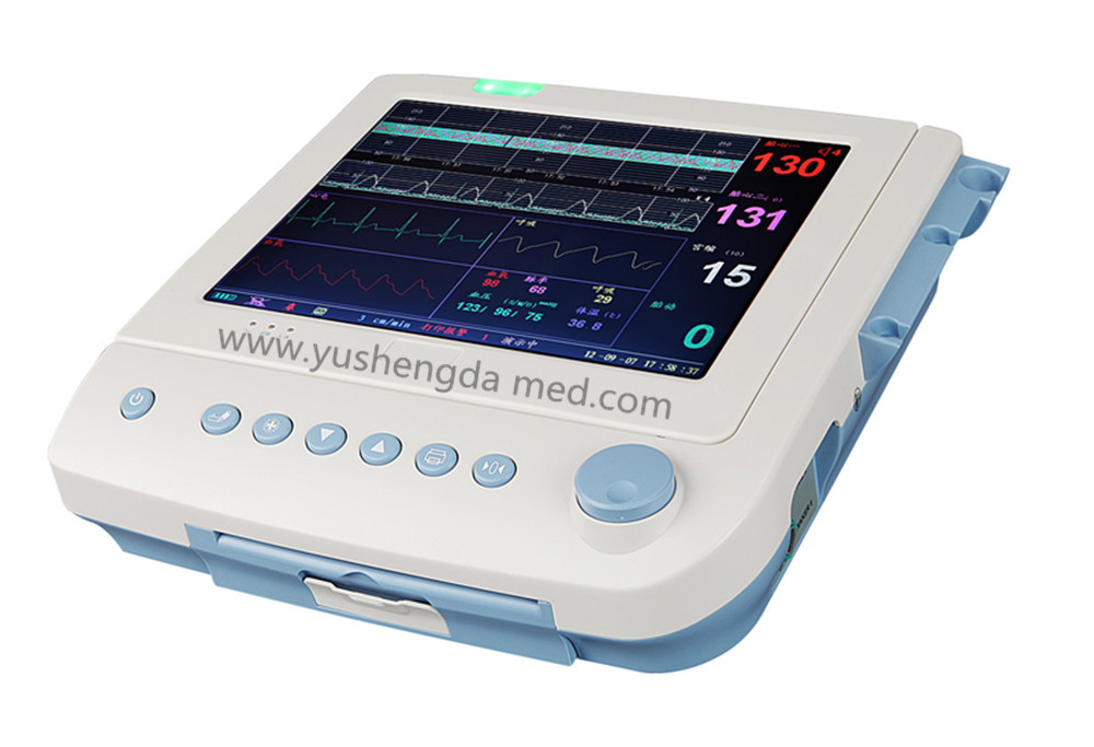
Although the evidence does not support this type of technology being used in low-risk pregnancies, EFM has become a standard of care in childbirth practice for women in the United States.Īpproximately 2 out of 1,000 children have cerebral palsy with the main risk factors for cerebral palsy being low birth weight, intrauterine infections, and multiple gestations.įACTORS INFLUENCING FETAL HEART RATE CHANGESįetal monitoring is used to assess the adequacy of fetal oxygenation during labor ( ACOG, 2009) with the goal being to prevent metabolic acidemia. By 1978, EFM was in routine use in one-half of all labors ( Williams & Hawes, 1979) in 2002, 85% of all women were assessed with EFM ( Martin et al., 2003).

Over time, the cardiography machine (EFM) became smaller and less bulky, which allowed this technology to fit at the bedside more easily and be used. Hon from Yale University was able to identify causes of bradycardia leading to fetal distress by monitoring the fetal heart rate continuously from the maternal abdomen (Hon & Lee, 1963).Ĭontinuous EFM was embraced by the obstetric community, including nursing ( Sandelowski, 2000), even though clinical trials did not show evidence supporting its use in low-risk women when compared to IA ( Banta & Thacker, 1979 Dixon, 1981 Haverkamp, Thompson, McFee, & Cetrulo, 1976). The first fetal electrocardiogram (EKG) recording was in 1906 ( Freeman & Garite, 1981) 50 years later, Dr. At the time, evaluating a fetus was primarily accomplished by putting one’s ear to the maternal abdomen, or using a Laennec instrument (cylindrical in shape and similar to the Pinard) to auscultate the fetal heart rate ( Freeman & Garite, 1981). The purpose of this article is to review the history of fetal monitoring address factors influencing the fetal heart rate during labor report on the different types of fetal monitoring available discuss barriers identified in the literature inhibiting the implementation of fetal monitoring choice in childbirth and provide suggestions to incorporate evidence-based material into childbirth classes.Īlthough the fetal heart sound was first described in a poem in the 1600s, it was not until the mid-1800s that abnormal fetal heart rates were associated with fetal distress signifying the need for a forceps intervention ( Freeman & Garite, 1981).

Many barriers exist preventing nurses from implementing IA during the intrapartum period ( Graham, Logan, Davies, & Nimrod, 2004 Lewis & Rowe, 2004 Rattray, Flowers, Miles, & Clarke, 2011 Regan & Liaschenko, 2007 Sleutel, Schultz, & Wyble, 2007). There is limited research exploring a woman’s ability to give informed choice regarding which method of fetal monitoring to use ( Hindley et al., 2008 O’Cathain, Thomas, Walters, Nicholl, & Kirkham, 2002). IA is a safe and acceptable fetal monitoring method that is recommended during labor with low-risk pregnancies ( ACOG, 2009 Anderson, 1994 Association of Women’s Health and Obstetric and Neonatal Nurses, 2008 National Institute of Clinical Excellence, 2007 The Royal Australian and New Zealand College of Obstetricians and Gynaecologists, 2009 United States Preventative Services Task Force, 1996 World Health Organization, 1996). An alternative option for healthy women with uncomplicated pregnancies is intermittent auscultation (IA).

Intermittent auscultation is a safe and acceptable fetal monitoring method that is recommended during labor with low-risk pregnancies.Ĭontinuous EFM is associated with many known medical risks to women, without providing any benefit to the fetus in low-risk pregnancies ( Alfirevic, Devane, & Gyte, 2006 ACOG, 2009). For AWHONN guidelines for Fetal Monitoring see:


 0 kommentar(er)
